HOURS: Mon–Sat

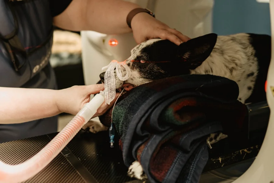
When something is wrong with your pet and the answer is not obvious, the next step is usually a picture. For decades that picture was an X-ray. Today we can go further.
Computed tomography, better known as CT, is an advanced imaging technique that uses X-rays to create cross-sectional images of the body. It enables detailed visualization of internal structures in animals, aiding both diagnosis and treatment planning.
That difference matters because CT offers more detailed images than X-ray. It has a wide range of applications in veterinary medicine, from dentistry to neurology to cancer care, and it can show a veterinarian exactly where a problem is and how far it extends.
For pet families in Kennewick, WA, and surrounding areas, understanding what CT can do makes it easier to say yes when your veterinarian recommends one.
What Is a CT Scan?
A CT scan is an advanced imaging technique that uses X-rays to create cross-sectional images of the body.
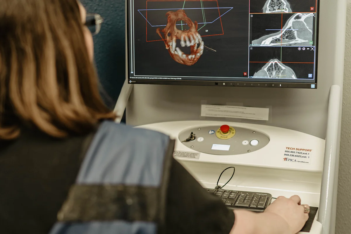
Rather than producing one flat picture, CT captures the body in slices. Those slices are reconstructed by a computer into a three-dimensional image, which allows detailed visualization of internal structures and supports both diagnosis and treatment planning.
How a CT Scan Works
The process is straightforward, and it happens while your pet is asleep.
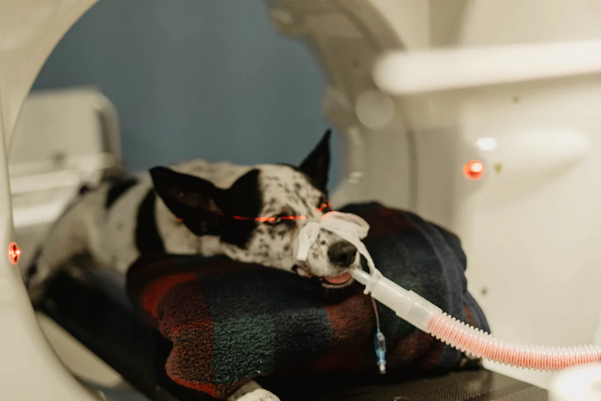
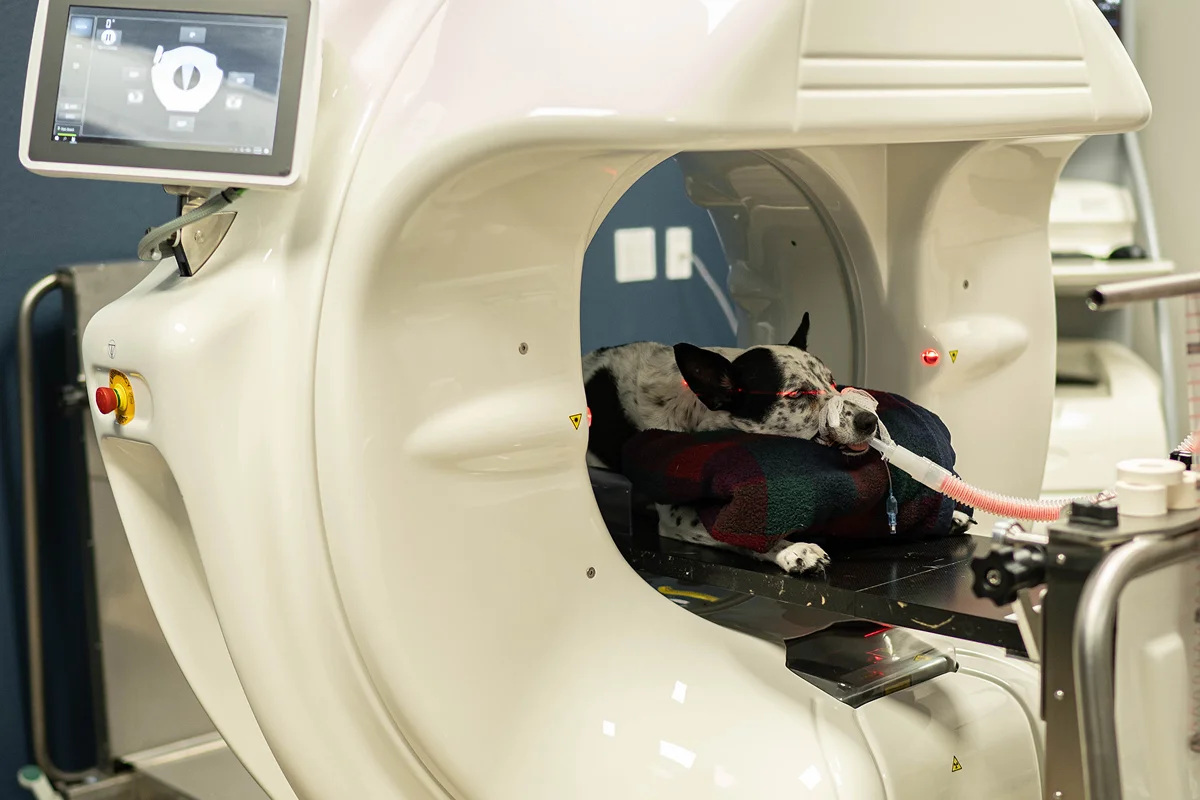
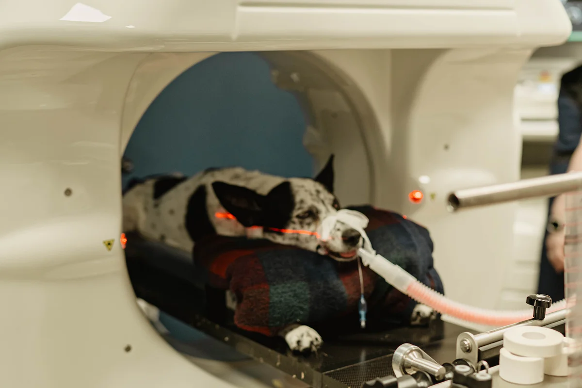
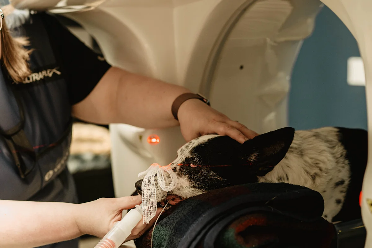
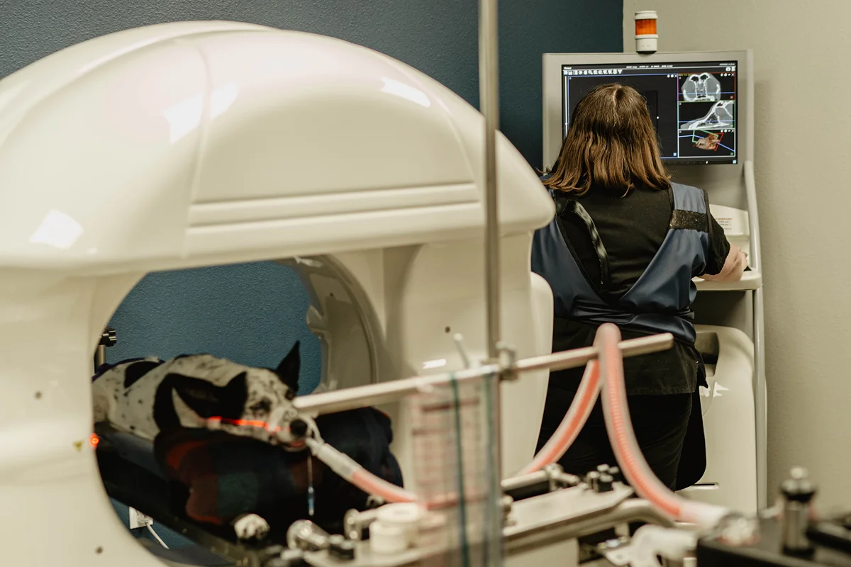
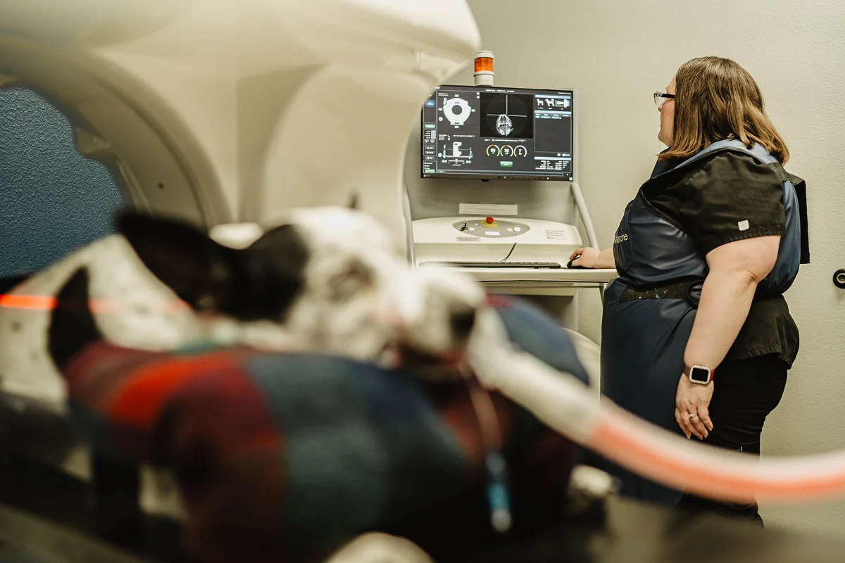
- Your pet is put under heavy sedation or general anesthesia to minimize movement.
- An X-ray tube, called a gantry, rotates around the body while detectors collect the data.
- That data is sent to a computer, which reconstructs it as a three-dimensional image.
- Contrast may be used to enhance the visibility of blood vessels and soft tissues.
Because the patient has to stay completely still for clear images, sedation is not optional. It is what makes the scan possible.
Advantages of Using CT
CT earns its place in a veterinary hospital for several reasons:
- High-resolution imaging of bones, organs and soft tissue
- Non-invasive, and images can typically be obtained quickly
- Useful for complex cases
- Allows for monitoring of treatment progress
- In dentistry, does not create issues with foreshortening or elongation
That last point deserves attention. On a standard dental radiograph, tooth roots can appear shorter or longer than they really are, which makes them difficult to assess. CT removes that distortion.
Common Applications for CT Imaging
CT is used across several areas of veterinary medicine:
- Dentistry — tooth root evaluation
- Neurology — brain tumors
- Orthopedics — fractures and joint abnormalities
- Oncology — tumor detection
- Cardiology — vascular anomalies
- Fluoroscopy studies — real-time evaluation of swallowing and esophageal function, helping diagnose conditions such as strictures, megaesophagus, and other esophageal disorders
What to Consider Before a CT Scan
CT is a powerful tool, and like any tool it has limitations worth discussing with your veterinarian:
- Cost. Because patients need to be sedated or anesthetized for the procedure, there is greater cost than for radiographs.
- Sedation or anesthesia is required. Every patient needs it in order to hold still.
None of these rule CT out. They are the reason your veterinarian will recommend it when the information it provides is worth having, rather than as a routine first step.
Build a Lasting Partnership with Horse Heaven Hills
Advanced imaging works best in the hands of a team that already knows your pet. At Horse Heaven Hills, our veterinarians use CT as one part of a larger picture that includes your pet’s history, exam findings and other diagnostic testing.
CT is a very valuable tool in veterinary medicine with a multitude of applications, including dentistry, neurology, oncology, orthopedics, and advanced fluoroscopy studies. These real-time imaging studies can help evaluate swallowing and esophageal function when conditions such as strictures or megaesophagus are suspected. When your veterinarian recommends advanced imaging, it is because a clearer picture can help guide the next steps in your pet’s care.
Talk to Us About Advanced Imaging for Your Pet
If you have questions about CT imaging, or your veterinarian has mentioned a scan and you would like to understand it better, contact Horse Heaven Hills. Our team proudly serves families throughout Kennewick, the Tri-Cities, and surrounding communities with advanced diagnostic imaging and same-day care.
Book Online | Call: (509) 581-0647
FAQs
Q1: What is a CT scan for pets?
A: Computed tomography is an advanced imaging technique that uses X-rays to create cross-sectional images of the body, enabling detailed visualization of internal structures to aid diagnosis and treatment planning.
Q2: Does my pet need to be sedated for a CT scan?
A: Yes. Patients are placed under heavy sedation or general anesthesia to minimize movement, because clear cross-sectional images require the patient to stay completely still.
Q3: How is a CT scan better than an X-ray?
A: CT offers more detailed images than X-ray, with high-resolution imaging of bones, organs and soft tissue. In dentistry it also avoids the foreshortening and elongation that affect standard radiographs.
Q4: What conditions can a CT scan help diagnose?
A: Common applications include tooth root evaluation in dentistry, brain tumors in neurology, fractures and joint abnormalities in orthopedics, tumor detection in oncology, and vascular anomalies in cardiology.
Q5: Why does a CT scan cost more than X-rays?
A: Because patients need to be sedated or anesthetized for the procedure, there is greater cost than for radiographs.
Q6: What is contrast used for during a CT scan?
A: Contrast is used to enhance the visibility of blood vessels and soft tissues, making certain structures
easier to evaluate on the reconstructed images.