HOURS: Mon–Sat

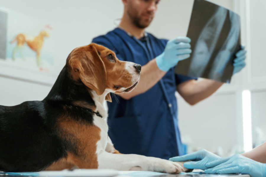
When a dog develops a persistent cough or a cat begins breathing differently, it is natural to wonder what is happening inside their body. A chest X-ray is one of the tools veterinarians use to investigate problems involving the lungs, heart, airways, and chest cavity. It creates images of structures inside the chest without surgery and can help identify changes that are not visible during a physical examination alone. A chest X-ray for dogs and cats may also be recommended after an injury or as part of an investigation into unexplained symptoms. At Horse Heaven Hills Pet Urgent Care and Veterinary Center in Kennewick, WA, pet owners can learn why imaging may be recommended and what the results can help a veterinarian understand. This guide explains what chest X-rays show, how the procedure works, and what happens afterward.
What Can a Chest X-Ray Show in Dogs and Cats?
A chest X-ray can help veterinarians assess the lungs, heart, airways, ribs, and space around the lungs. It may reveal signs of infection, fluid buildup, an enlarged heart, masses, certain injuries, or other changes that help explain a pet’s symptoms. The images provide valuable information, but they do not always identify the exact cause of a problem on their own.
Chest X-rays are also called thoracic radiographs. They use a small amount of radiation to create images of the structures inside the chest. Different tissues appear in different shades, allowing a veterinarian to compare the size, shape, and appearance of organs and surrounding areas.
Depending on the reason for the examination, a chest X-ray may help evaluate:
- The lungs: Changes that may be associated with inflammation, infection, fluid, or masses.
- The heart: Its general size and shape, along with signs that may suggest certain heart conditions.
- The airways: Changes in the trachea and larger airways that may contribute to coughing or breathing problems.
- The chest cavity: Fluid or air in spaces where they should not normally collect.
- The bones: Some fractures or other changes involving the ribs and nearby structures.
- The esophagus and surrounding areas: Certain abnormalities may be visible depending on their size and location.
The findings are interpreted alongside your pet’s symptoms, physical examination, medical history, and sometimes laboratory results. For example, a cough may be caused by a problem in the airways, lungs, heart, or another part of the body. Imaging helps narrow the possibilities.
A normal-looking X-ray does not rule out every disease. Some conditions are difficult to see on radiographs, especially in early stages or when changes are subtle. Your veterinarian may recommend additional testing if the images do not fully explain your pet’s symptoms.
Why Would a Dog or Cat Need a Chest X-Ray?
Veterinarians recommend chest imaging when they need more information about symptoms, an injury, or a possible condition affecting the chest. The decision depends on your pet’s signs, age, medical history, and examination findings.
Common reasons for a chest X-ray include:
- A persistent or unexplained cough.
- Faster or more difficult breathing.
- Unusual breathing sounds.
- Reduced stamina or exercise tolerance.
- Suspected heart or lung disease.
- Chest trauma after a fall or accident.
- Possible fluid or air around the lungs.
- Follow-up of a known condition.
- Investigation of certain masses or suspected cancer.
- Assessment before some procedures when imaging is medically appropriate.
Coughing and breathing changes
Coughing can develop for many reasons. Dogs may cough because of airway irritation, infection, heart disease, or other conditions. Cats can also develop respiratory problems that affect their lungs or airways, and some cats show subtle signs rather than an obvious cough.
A chest X-ray may help identify a pattern of changes that supports one possible cause over another. However, the image must be interpreted with the examination and other findings because different diseases can produce similar appearances.
Injury or suspected chest damage
After an accident, imaging may help identify rib fractures, changes in the lungs, or air or fluid around the lungs. Not every injury is visible on an X-ray, so the veterinarian may also assess pain, breathing, circulation, and other signs.
If your pet has difficulty breathing after an injury, seek urgent veterinary care rather than waiting for a routine appointment.
Investigation of other health concerns
Sometimes, chest imaging is recommended when a pet has unexplained weakness, weight loss, or other signs that may be connected to a condition inside the chest. It may also be used to investigate certain masses or evaluate whether a known illness has affected other areas.
Not every pet with a cough or other symptom needs an X-ray. The veterinarian will decide whether imaging is likely to provide useful information and whether another test should come first.
What Can a Chest X-Ray Reveal About a Dog’s Lungs?
A chest X-ray for dogs can show changes in the lungs that may help explain coughing, breathing problems, or reduced activity. Healthy lungs contain air, which usually makes them appear relatively dark on an X-ray. When inflammation, fluid, tissue changes, or other material affects the lungs, parts of the image may appear whiter than expected.
Possible findings include:
- Changes associated with pneumonia: Inflammation and fluid within the lungs can create patterns that support a diagnosis when combined with symptoms and other tests.
- Airway changes: Thickening or other changes around the airways may be visible in some respiratory conditions.
- Fluid in or around the lungs: The location of the fluid can help guide the veterinarian’s investigation.
- Masses or nodules: Some abnormal growths can be seen, although X-rays may not determine exactly what a mass is.
- Changes following an injury: Imaging may reveal certain lung injuries or air around the lungs.
- Abnormal lung patterns: These can help the veterinarian decide whether further testing is needed.
Can an X-ray diagnose pneumonia?
A chest X-ray can provide evidence that supports pneumonia, but it may not always confirm the cause. The veterinarian considers symptoms such as coughing, fever, reduced appetite, and breathing changes, along with the appearance of the lungs.
Additional tests may be recommended if the findings are unclear or if identifying the cause would change treatment. The choice depends on how sick the dog is and which conditions are most likely.
What if the X-ray looks normal?
A normal chest X-ray can be reassuring, but it does not rule out every respiratory condition. Some problems may be too subtle to appear on the images, or they may involve structures that are better assessed with other tests.
If your dog continues coughing or breathing abnormally, the veterinarian may recommend further examination, repeat imaging, laboratory testing, or another diagnostic approach. Do not assume ongoing symptoms are harmless simply because an earlier image looked normal.
The purpose of the X-ray is to add information to the overall assessment. It is one part of the diagnostic process, not a replacement for the physical examination or clinical judgment.
What Can a Chest X-Ray Reveal About a Cat’s Heart and Lungs?
A chest X-ray for cats can help veterinarians assess the appearance of the lungs, heart, airways, and surrounding chest structures. It is often considered when a cat has breathing changes, persistent coughing, unexplained weakness, or a suspected heart or lung condition.
Cats can hide signs of illness, so even small changes in their normal behavior may matter. A cat that is quieter than usual, eats less, or seems to tire easily may need an examination, particularly if breathing also looks different.
Evaluating the lungs and airways
Chest images may show changes that support conditions involving inflammation, infection, asthma-like airway disease, fluid, or other lung problems. However, these conditions can overlap in appearance, and a veterinarian may need additional information to determine the likely cause.
The images may also help identify fluid or air around the lungs. These findings can be particularly important when a cat is breathing rapidly or struggling to breathe.
Assessing the heart
A chest X-ray provides information about the heart’s general size and shape and may show changes associated with some heart conditions. It can also help identify certain effects that heart disease may have on the lungs or surrounding chest structures.
However, a chest X-ray cannot fully assess how well the heart muscle contracts or how blood moves through the heart. An echocardiogram, which uses ultrasound to examine the heart, may be recommended when a more detailed assessment is needed.
Some cats with heart disease have few obvious signs until the condition becomes more serious. If your cat develops rapid or labored breathing, open-mouth breathing, collapse, or sudden weakness, seek immediate veterinary care.
Why additional tests may be needed
A veterinarian may recommend blood tests, ultrasound, or other diagnostic procedures depending on the symptoms and X-ray findings. The right combination of tests helps distinguish between possible causes and guides treatment.
Avoid trying to diagnose a cat’s breathing problem based on an image or a symptom alone. A veterinarian can interpret the findings in the context of the cat’s age, history, examination, and current condition.
How Is a Veterinary Chest X-Ray Performed?
A veterinary chest X-ray is usually a short imaging procedure, although the time required depends on your pet’s condition and how easily the images can be obtained. The veterinary team positions your pet so the images show the chest clearly while keeping handling as gentle as possible.
Here is what pet owners can generally expect:
- Initial assessment: The veterinarian examines your pet and decides whether chest imaging is appropriate.
- Positioning: A trained team member positions your pet on an imaging table or another suitable surface. The position depends on the views needed and your pet’s comfort.
- Image capture: The X-ray machine takes images from selected angles. More than one view is often needed because organs and structures overlap in a single image.
- Image review: The veterinarian examines the images for abnormalities and compares the findings with the symptoms and physical examination.
- Discussion of results: The veterinarian explains what the images show, what they may mean, and whether additional tests or treatment are recommended.
Does my pet need sedation?
Many pets can have chest X-rays without sedation. However, a pet that is frightened, painful, or unable to remain in the needed position may require additional support. In selected cases, the veterinarian may recommend sedation to reduce stress and help obtain useful images.
The decision depends on your pet’s health, behavior, and the reason for imaging. If sedation is being considered, the veterinary team can explain the benefits, risks, and any preparation needed.
Is the procedure painful?
Taking an X-ray does not usually cause pain. However, positioning may be uncomfortable for a pet with an injury or breathing difficulty. The team should consider your pet’s condition when deciding how to handle and position them.
If your pet is struggling to breathe, tell the veterinary team immediately and follow their instructions. Stabilizing a pet’s breathing may take priority over routine imaging.
Hypothetical case example
A middle-aged dog develops a persistent cough and tires more quickly during walks. After examining the dog and listening to the heart and lungs, the veterinarian recommends chest X-rays to look for changes involving the airways, lung tissue, or heart. The images show an abnormal pattern that helps guide the next diagnostic steps, but the veterinarian explains that additional information is needed before confirming the cause. This hypothetical example illustrates how imaging can narrow the possibilities without always providing a final diagnosis on its own.
The veterinary team will explain what to expect based on your pet’s condition. If you have questions about handling, sedation, or the purpose of the images, ask before the procedure begins.
What Happens After a Pet’s Chest X-Ray?
After the images are taken, the veterinarian reviews them and explains the findings. Sometimes, the results provide a likely explanation for the symptoms. In other cases, the images identify an area of concern but cannot determine its exact cause.
The next step depends on what the X-ray shows and how your pet is feeling. Your veterinarian may recommend:
- Treatment based on the findings and examination.
- Blood work or other laboratory tests.
- Ultrasound or another type of imaging.
- Additional tests to investigate a suspected condition.
- Repeat X-rays to monitor changes over time.
- Referral for further evaluation if the condition requires it.
A pet with significant breathing difficulty may need treatment and stabilization before additional testing can be completed. A pet that is stable may have more time for a step-by-step diagnostic plan.
Ask your veterinarian to explain the main findings in plain language. Useful questions include:
- What abnormalities, if any, were seen?
- Do the images explain my pet’s symptoms?
- Is another test needed to confirm the suspected cause?
- What signs should I watch for at home?
- When should my pet return for a follow-up?
If your pet returns home after the examination, follow the instructions provided by the veterinary team. Monitor appetite, activity, coughing, and breathing as advised. Do not change prescribed medication or stop treatment because your pet seems better without checking with the veterinarian.
Seek prompt care if symptoms worsen. Breathing difficulty, blue or gray gums, collapse, or open-mouth breathing in a cat should be treated as urgent signs that require immediate veterinary attention.
Are Chest X-Rays Safe for Dogs and Cats?
Chest X-rays are widely used in veterinary medicine because they provide useful information without requiring surgery. The procedure uses a controlled amount of radiation, and the veterinary team aims to obtain the necessary images while avoiding unnecessary exposure.
The benefits and limitations should be considered for each pet. When imaging is likely to help identify a serious problem or guide treatment, the information it provides may be important to the pet’s care.
The team may use positioning aids or other measures to reduce movement and improve image quality. Clear images can help avoid the need to repeat the procedure.
Some pets require special consideration. A dog with severe pain or a cat with breathing difficulty may not tolerate normal positioning. The veterinarian will decide how to balance the need for imaging with the pet’s comfort and stability.
It is also important to understand what an X-ray cannot show. Although it can reveal many changes in the chest, it does not provide the same detail as every other diagnostic method. Ultrasound, laboratory testing, or other imaging may be more useful for certain questions.
If your veterinarian recommends a chest X-ray, ask what they are looking for and how the result may change the treatment plan. Understanding the purpose of the test can make the process less stressful and help you make informed decisions about your pet’s care.
When Should I Seek Veterinary Care for Breathing Problems?
Breathing changes should always be taken seriously, especially if they appear suddenly or worsen quickly. A chest X-ray may be useful during the investigation, but a veterinarian must first assess how stable your pet is.
Contact a veterinarian promptly if your pet has a new or persistent cough, unusual breathing sounds, reduced stamina, or a noticeable change in normal activity. These signs may have several causes, and an examination can help determine whether imaging is appropriate.
Seek immediate veterinary care if your pet has:
- Labored or unusually rapid breathing.
- Open-mouth breathing, particularly in cats.
- Blue, gray, or very pale gums.
- Collapse or sudden severe weakness.
- Significant breathing difficulty after an injury.
- A posture suggesting that breathing is difficult, such as stretching the neck to breathe.
Keep your pet as calm as possible while arranging care. Avoid forcing food, water, or medication if breathing is difficult, and do not delay seeking help to try home remedies.
Not every cough requires a chest X-ray, and not every abnormal X-ray requires the same treatment. The veterinarian will consider your pet’s symptoms, examination, and test results before recommending the next step.
Horse Heaven Hills Pet Urgent Care and Veterinary Center can assess concerning symptoms and help determine whether chest imaging or other testing is appropriate. If your pet’s breathing is severely affected, seek immediate veterinary attention rather than waiting for a routine visit.
Conclusion
A chest X-ray can help veterinarians evaluate a dog’s or cat’s lungs, heart, airways, chest cavity, and certain injuries. It may reveal changes associated with infection, fluid, masses, or other conditions, but the findings do not always provide a final diagnosis. Your veterinarian may recommend additional tests to understand the cause and choose appropriate treatment.
If your pet has a persistent cough, unexplained breathing changes, or a suspected chest injury, ask whether imaging may help. Horse Heaven Hills Pet Urgent Care and Veterinary Center in Kennewick, WA, can help you understand the role of chest X-rays in evaluating your pet’s health. Seek immediate care for labored breathing, collapse, or other severe symptoms, because timely assessment can be critical.
FAQs
Q1: What can a chest X-ray detect in a dog or cat?
A: Chest X-rays can show changes involving the lungs, heart, airways, chest cavity, and some bones. They may help identify signs of infection, fluid buildup, certain masses, or injuries. Additional tests may be needed to determine the exact cause.
Q2: Does my pet need to be sedated for a chest X-ray?
A: Many pets can have chest X-rays without sedation. Sedation may be considered if a pet is very anxious, painful, or unable to remain in position safely. Your veterinarian will decide based on your pet’s condition and the images needed.
Q3: Can a chest X-ray diagnose heart disease in pets?
A: A chest X-ray can show the heart’s general size and shape and reveal some changes associated with heart disease. It cannot assess every aspect of heart function, so an echocardiogram or other testing may be recommended.
Q4: How long does a veterinary chest X-ray take?
A: Taking the images is often relatively quick, but the full visit may take longer because of the examination, positioning, image review, and discussion of results. The time depends on your pet’s condition and whether additional tests are needed.
Q5: When should my dog or cat have a chest X-ray?
A: A veterinarian may recommend chest imaging for persistent coughing, breathing changes, suspected heart or lung disease, chest injuries, or certain unexplained symptoms. Labored breathing, collapse, or open-mouth breathing in a cat requires immediate veterinary attention rather than waiting for a routine appointment.